
INTRODUCTION
Clinical Overview
This clinical resource presents a comprehensive overview of endodontic procedures for pediatric patients. The guide is structured into specialized sections designed for orderly clinical navigation and procedural clarity.
- Primary Teeth: Clinical coverage ranging from protective pulp therapy to comprehensive root canal management.
- Immature Permanent Teeth: Advanced endodontic treatments encompassing both apexogenesis and apexification protocols.
- Traumatic Dental Injuries: Standardized management for fractures, luxations, and avulsions across all pediatric dental types.
Professional Biography
Michael L. Sosnay, DDS is an Endodontic specialist practicing in New York City since 1999. He received his Doctorate of Dental Surgery in 1995 from New York University Dental School, where he was an NYU Dean’s Dental Merit Scholar. Dr. Sosnay holds a dual specialty degree in Endodontics and Pediatric Special Care dentistry. He received his Post-Doctoral Degree in Endodontics in 1999 and served as Director of Endodontics and Associate Clinical Professor at the Rose F. Kennedy Center – Jacobi Hospital/Albert Einstein.
Dr. Sosnay has lectured extensively on endodontic theory, diagnosis, and treatment modalities, as well as pediatric endodontics and trauma. He is a member of the American Dental Association, New York State Dental Association, New York County Dental Society, American Association of Endodontists, and the Special Care Dentistry Association.
- Dental Traumatology References:
Evidence-based protocols for clinical diagnosis, active management, and long-term follow-up:
• AAE: Guidelines developed with IADT (aae.org)
• IADT: Published trauma guidelines (iadt-dentaltrauma.org)
• AAPD: Guidance for primary teeth trauma (aapd.org)
• Dental Trauma Guide: Foundational reference (dentaltraumaguide.org)
Resources & Guidelines
- Pulp Therapy References:
Clinical references for primary and immature permanent teeth:
• AAPD: Clinical Practice Guidelines (aapd.org)
• AAE & AAPD Joint Symposium: Consensus recommendations on pediatric pulp management.
Disclaimer: These clinical guidelines provide evidence-based information for healthcare providers managing pediatric dental injuries. Clinical judgment must remain paramount, dictated by the specific conditions of each traumatic situation. The IADT and related associations maximize treatment success via these procedures but do not guarantee outcomes.
Primary Teeth – Pulp Therapy
SPACE MAINTENANCE RULE
If the "E" tooth (primary second molar) is lost early, space maintenance is required to allow the premolar to erupt properly.
If the "D" tooth (primary first molar) is lost early, it depends on eruption status. If the 6-year molar has fully erupted, space maintenance is not necessary. If it has not erupted, space maintenance is required, as eruption can drive the "E" tooth mesially and block the premolar from erupting.
1. Problem: Deep Caries
Treatment: Protective Liner
- Excavate decay.
- Place a liner such as CaOH, dentin bonding agent, or glass ionomer.
- Cover with a permanent restoration.

2. Problem: Small Carious Pulpal Exposure with No Significant Pain
Treatment: Indirect Pulp Cap
- Excavate gross caries.
- Leave caries directly over the pulp chamber to avoid pulpal exposure.
- Cover with a biocompatible material such as CaOH or glass ionomer to stimulate healing.
- Place a permanent restoration.

3. Problem: Pinpoint Pulpal Exposure During Cavity Preparation or Trauma
Treatment: Direct Pulp Cap
- Cover pulp tissue with a biocompatible material such as CaOH or MTA.
- Place a permanent restoration.
.webp)
4. Problem: Large Carious Pulpal Exposure with No Significant Pain
Treatment: Pulpotomy
- Remove pulp tissue in the pulp chamber.
- Apply a wet cotton pellet with sodium hypochlorite to control bleeding.
- Fill chamber with Buckley’s solution, glutaraldehyde with CaOH, or MTA (most successful).
- Place a permanent restoration.
- Cover with a stainless steel crown for long-term clinical success.
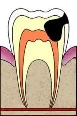
5. Problem: Non-Vital Pulp Tissue, Furcal Bone Loss, or Irreversible Pulpitis Pain
Treatment: Pulpectomy
- Clean and fill the canals with a resorbable material such as KRI or Vitapex.
- These materials resorb naturally along with the roots.
- Place a permanent restoration.

6. Problem: No Permanent Successor and Necrotic Pulp Tissue or Irreversible Pulpitis Pain
Treatment: Conventional Root Canal Therapy
- Perform conventional RCT protocols.
- Fill the canals with a non-resorbable material such as gutta-percha with sealer or MTA.
- Place a final permanent restoration.

Objective: Preserve pulp tissue.
Reason: Primary teeth with vital pulp tissue have a better prognosis and a greater chance of exfoliating on time, helping to avoid space maintenance and orthodontic complications.
Immature Permanent Teeth
Objective: Preserve pulp tissue.
Reason: Vital pulp tissue is necessary for immature teeth to complete root development, improving long-term prognosis.
1. Problem: Deep caries with no pulpal exposure and no significant pain.
Treatment: PROTECTIVE LINER — excavate decay and place a thin protective liner like CaOH, dentin bonding agent, or glass ionomer over the pulp chamber to protect the pulp, and then place a permanent restoration on top.

2. Problem: Small carious pulpal exposure and no significant pain.
Treatment: INDIRECT PULP CAP — excavate the gross caries, leaving a carious mass over the pulp to avoid pulp exposure; then place a protective liner like glass ionomer over the remaining caries and cover with a permanent restoration. Note: For small carious pulpal exposures, indirect pulp capping is the preferred method.
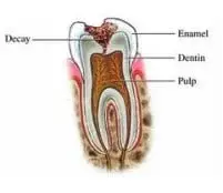
3. Problem: Pinpoint pulpal exposure encountered during decay excavation or traumatic injury and normal pulp tissue (e.g., asymptomatic or reversible pulpitis).
Treatment: DIRECT PULP CAP — the pulp tissue is covered with MTA, which is covered with a wet cotton pellet (to allow the MTA to set) and temporized. The patient returns to have the temporary restoration and cotton removed and a permanent filling is placed.
Note: For small carious pulpal exposure, the indirect pulp cap is the preferred method.

4. Problem: Large carious pulpal exposure and normal pulp tissue (e.g., asymptomatic or reversible pulpitis).
- Treatment: PARTIAL PULPOTOMY (CVEK) — remove 1-3 mm of coronal pulp tissue (with a high-speed diamond bur with coolant) beyond the caries to reach healthy pulp tissue; dab remaining pulp tissue with a sodium hypochlorite cotton pellet to control bleeding; cover the remaining pulp tissue with MTA; cover the MTA with a wet cotton and temporary filling. The patient returns to have the temporary restoration and cotton removed and a permanent restoration placed.
- Treatment: FULL PULPOTOMY — remove all pulp tissue from the chamber; control bleeding with sodium hypochlorite cotton pellet; fill the pulp chamber with MTA and cover with wet cotton and temporize. The patient returns for the removal of the temporary restoration and cotton, and a permanent restoration is placed.

Note: All four of the above problems can be considered apexogenesis, as they promote apical root development by aiming to preserve vital pulp tissue.
5. Problem: Apexified tooth (i.e., closed apices) with necrotic pulp tissue or irreversible pulpitis.
Treatment: CONVENTIONAL RCT.

6. Problem: Incompletely developed tooth with open apices, with necrotic pulp tissue or irreversible pulpitis.
Treatment: UNCONVENTIONAL RCT — remove all the pulp tissue from the chamber and canals and fill them with CaOH for 2–4 weeks to disinfect the canal space. Then bring the patient back to remove the CaOH, place an MTA apical seal, and backfill with gutta-percha.

Note: On a tooth with a wide-open apex (e.g., #8), you can place CollaCote (collagen) just past the apex in order to have something firm to pack the MTA up against, avoiding extrusion.
Clinical Resources
If the "E" tooth (primary second molar) is lost early, space maintenance is required to allow the premolar to erupt properly.
Refer to IADT guidelines for traumatic dental injuries and AAPD protocols for pulp therapy. Learn more at www.iadt-dentaltrauma.org and www.dentaltraumaguide.org.
Traumatic Dental Injuries
Clinical protocols for the diagnosis and management of dental trauma.
CLINICAL FRAMEWORK
Each condition below follows a standardized format:
- Diagnosis: Clinical identification of the injury.
- Treatment: Specific management protocols.
- Prognosis: Expected long-term outcome.
- Patient Instructions: Clinical guidance for home care.
When "Monitor" is indicated, refer to the trauma follow-up protocol outlined in the Charts section.
I. Permanent Teeth Management
1. Problem: Concussion
- Diagnosis: Tooth is tender to touch.
- Treatment: MONITOR.
- Prognosis: Pulp complications are rare and root resorption is very rare.
- Patient Instructions: Soft food for 1 week, good oral hygiene, and chlorhexidine rinse for 2 weeks.
2. Problem: Subluxation
- Treatment: Flexible splint (0.016" or 0.4 mm) for 2 weeks is optional, then MONITOR.
- Prognosis: Pulp complications are rare and root resorption is very rare.
- Patient Instructions: Soft diet for 1 week, good oral hygiene, and chlorhexidine rinse for 2 weeks.
3. Problem: Lateral Luxation
- Treatment: Rinse area with saline, REPOSITION the tooth with your fingers or a forceps: Pull the tooth coronally at first to free the tooth from its incorrect bony locked position, and then back apically into its proper position. Flexible splint for 2–4 weeks.
- Open Apex: Can revascularize, so MONITOR. If pulp devitalizes, options include CaOH traditional apexification, immediate RCT with MTA seal of apex, or regenerative pulp therapy.
- Closed Apex: Pulp necrosis is common, so conventional RCT can be initiated before splint removal or MONITOR.
- Prognosis:
- Open Apex: Guarded pulpal prognosis.
- Closed Apex: Poorer pulpal prognosis.
- Root resorption is rare.
- Patient Instructions: Soft diet for 1 week, good oral hygiene, and chlorhexidine rinse for 2 weeks.
4. Problem: Extrusion Luxation
- Treatment: Rinse the area with saline, REPOSITION, and flexible splint for 2 weeks.
- Open Apex: Can revascularize, so MONITOR. If pulp devitalizes, see treatment options listed in Case 3.
- Closed Apex: Pulp necrosis is common, so conventional RCT can be initiated right before splint removal, or MONITOR.
- Prognosis:
- Open Apex: Guarded pulpal prognosis.
- Closed Apex: Poorer pulpal prognosis.
- Root resorption is rare.
- Patient Instructions: Soft diet for 1 week, good oral hygiene, and chlorhexidine rinse for 2 weeks.
5. Problem: Intrusion Luxation
- Treatment: REPOSITION.
- Open Apex:
- 0–7 mm intruded: Allow 3 weeks to re-erupt. If it doesn't, initiate orthodontic repositioning.
- 7 mm or more intruded: Immediately reposition orthodontically or surgically.
- Revascularization is possible in both cases, so MONITOR.
- Closed Apex:
- 0–3 mm: Allow 3 weeks to re-erupt. If not, reposition orthodontically or surgically. Then RCT (CaOH 4 weeks prior to fill) as the pulp will likely necrose.
- 3–7 mm: Reposition orthodontically or surgically and flexible splint for 2 weeks, then RCT.
- 7 mm or more: Reposition surgically and flexible splint 2–4 weeks, then RCT.
- Prognosis:
- Open Apex: Guarded pulpal prognosis.
- Closed Apex: Poor pulpal prognosis.
- Root Resorption: Guarded prognosis, as infection and ankylosis-related resorption are frequent findings.
- Patient Instructions: Soft diet for 1 week, good oral hygiene, and chlorhexidine rinse for 2 weeks.
6. Problem: Infraction
- Treatment: Etch and seal with resin to prevent discoloration. No need to monitor.
7. Problem: Uncomplicated Fracture
- Treatment: Three options include: glass ionomer bandage, permanent composite BONDING, or bonding the fragment back if available. Follow-up at 8 weeks and 1 year.
- Prognosis: Pulp complications and root resorption are rare.
- Patient Instructions: Soft diet for 1 week, good oral hygiene, and chlorhexidine rinse for 2 weeks.
8. Problem: Complicated Fracture
- Treatment:
- Open Apex: MTA PULP CAP or MTA PARTIAL PULPOTOMY (Cvek) and Restore. Research shows very high success rates for both. Follow-up at 8 weeks and 1 year.
- Closed Apex: Three options include: MTA PULP CAP, MTA PULPOTOMY, or conventional RCT. Follow the follow-up protocol listed in the charts section.
- Prognosis:
- Open Apex: Remaining pulp tissue has a good prognosis.
- Closed Apex: Remaining pulp tissue has a good prognosis.
- Root resorption is rare.
9. Problem: Crown Root Fracture
- Treatment: The primary goal is to create a situation where the tooth can be restored after fragment removal. EXPOSE THE SUBGINGIVAL FRACTURE SITE via gingivectomy, surgical exposure, orthodontic extrusion, or surgical extrusion.
- Uncomplicated (no pulp exposure): Remove fragment and restore.
- Complicated (pulp exposure):
- Open Apex: Perform MTA PULP CAP or MTA PULPOTOMY (apexogenesis). Follow-up at 8 weeks and 1 year.
- Closed Apex: Perform RCT and restore with a post.
- Prognosis:
- Uncomplicated: Both pulp and root have a good prognosis.
- Complicated:
- Open Apex: Remaining pulp tissue has a good prognosis.
- Closed Apex: The root has a good prognosis once RCT is performed.
- Root resorption is rare.
- Patient Instructions: Soft diet and soft brush.
10. Problem: Horizontal Root Fracture
- Treatment: REPOSITION the coronal fragment and stabilize with a flexible splint for 3–4 weeks and monitor. If at the CEJ, splint for 4 months and monitor. If RCT is indicated based on testing outcomes, cleaning and filling to the fracture site is suggested.
- Prognosis:
- Overall Prognosis: The further apical the fracture, the better the prognosis.
- Pulpal Prognosis: Depends on tooth maturity and degree of luxation. Less maturity and less displacement improve revascularization chances.
- Root Resorption Prognosis: Healing resorption or remodeling is common; pathological resorption is rare.
- Patient Instructions: Soft diet and soft brush.
- Biological Considerations: The coronal portion can be considered a luxated tooth with damage to neurovascular supply and PDL, while the apical portion remains relatively uninjured.
- Healing Types:
- Hard Tissue: New dentin and cementum bridge the fracture gap (immature teeth/young patients).
- Connective Tissue: PDL cells fill the gap (mature teeth).
- Granulation Tissue: Result of a necrotic pulp complex (necrotic teeth).
11. Problem: Alveolar Fracture
- Treatment: REPOSITION the fragment and stabilize with a flexible splint for 3–4 weeks, and MONITOR. Pulpal necrosis is a frequent finding, so RCT will probably be indicated. Treatment of choice for associated avulsion is Replantation. Site first aid: rinse, replant, bite on gauze. If cannot replant: store in milk, saliva, or saline.
- Prognosis: Poor pulpal prognosis due to frequent necrosis. Root resorption is rare.
- Patient Instructions: Soft diet and soft brush.
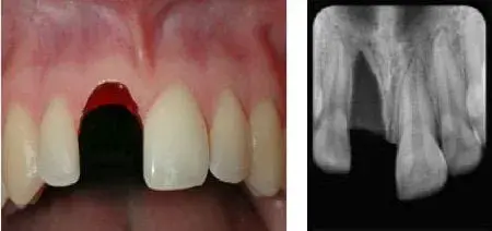
12. Problem: Avulsed - Replanted
- Treatment: Clean site with saline. Verify position with X-ray. Reposition up to 48 hours with mepivacaine. Non-rigid splint for 2 weeks (0.4 mm wire, avoid soft tissue). Administer antibiotics (Amoxicillin 250 mg TID for children). Monitor via follow-up protocol. Tetanus prophylaxis.
- Open Apex: Monitor for revascularization.
- Closed Apex: RCT in 2 weeks with CaOH.
- Prognosis:
- Pulpal Prognosis (Open Apex): Guarded.
- Root Resorption: Guarded.
- Patient Instructions: Good oral hygiene, soft diet, and chlorhexidine rinse.
- Biological Considerations: Revascularization and reinnervation take place from apical to coronal at a rate of 0.5 mm per day. Success depends on apical foramen size and absence of bacteria. Failure leads to pulp necrosis; success leads to canal obliteration. Repaired apical bone may initially appear as a PAR; monitor for bone regrowth if tooth remains vital.
13. Problem: Avulsed - In Storage
- Treatment: Clean root with saline. Use mepivacaine. Irrigate socket with saline to clear coagulum. Replant tooth with gentle pressure. Verify position with X-ray. Flexible splint for 1–2 weeks. Administer antibiotics (Amoxicillin 250 mg TID for children). Tetanus prophylaxis. Maintain good oral hygiene, soft diet, and chlorhexidine rinse.
- Open Apex: Monitor for revascularization.
- Closed Apex: RCT in 2 weeks with CaOH.
- Prognosis: Depends on extraoral time (Classifications: <15 min: PDL viable; <1 hr: PDL compromised; >1 hr: PDL not viable).
- Pulpal Prognosis (Open Apex): Guarded.
- Root Resorption: Guarded.
- Patient Instructions: Good oral hygiene, soft diet, and chlorhexidine rinse.
- Biological Considerations: Revascularization occurs at 0.55 mm/day from the apex, depending on foramen size and bacteria. Successful revascularization leads to canal obliteration. External resorption requires immediate RCT; change CaOH every 3 months if necessary. Conventional RCT is typically required for lateral luxation, intrusion, severe extrusion, and avulsion.
- Note: Rigid splints increase risk of ankylosis and resorption. Keep composite blobs away from soft tissue to prevent bacterial accumulation.
.webp)


Charts:
- MONITOR: Follow up chart for dental trauma
- Follow up intervals: 3 weeks, 6 weeks, 6 months, 1 year, once annually for the next 5 years.
- Follow up tests: percussion test, apical palpation test (look and feel for swelling), discoloration test, mobility test, pulpal vitality test, radiographic evaluation (look for periapical pathology and resorption)
- Root canal treatment is indicated for the following outcomes of follow up testing:
- Pulp tests non-vital. Diagnosis: Pulpal necrosis.
- Lingering pain to cold. Diagnosis: Irreversible pulpitis.
- X-ray shows periapical pathology and the pulp tests non-vital. Diagnosis: Chronic apical periodontitis.
- X-ray shows resorption. Diagnosis: Internal or external resorption, can take a CBT scan to determine which one.
Note: Transient or remodeling resorption, which is not pathological, but is part of healing, takes place in the few weeks following the trauma. But resorption that begins well after this point is pathological and requires treatment. - Discoloration
- Moderate to severe percussion sensitivity after 3 weeks. Diagnosis: Acute apical periodontitis.
- Treatment urgency chart:
- Acute: i.e. needs immediate attention: avulsions, alveolar fracture extrusion, lateral luxations, and root fractures.
- Subacute: i.e. delayed treatment is o.k: concussion, subluxation, intrusion, complicated crown fracture i.e. with pulp exposure.
- Resorption and healing:
- Repair related resorption: sometimes an area develops and then disappears during the healing process.
- Infection related resorption: bacteria stimulate the cementoclasts and that could destroy the tooth itself.
- Ankylosis: the PDL is injured in a certain area, so all new tissue growth comes from the bony socket and not the PDL, so the bone ends up growing right up against the root surface.
- Transient marginal bone resorption: bone breakdown in the coronal portion of the root, and it then regenerates.
II. Primary Teeth:
Problem:
Treatment:
1. Enamel fracture
Smooth sharp edges.
2. Enamel dentin fracture
Seal with glass ionomer or composite.
3. Crown fracture with pulp exposure
Preserve pulp with partial pulpotomy; seal pulp with MTA; composite on top.
4. Horizontal root fracture
If coronal displaced: splint or extract.
5. Alveolar fracture
Reposition displaced segment; splint; monitor.
6. Concussion: tender to touch
No treatment: observe.
7. Subluxation: mobility
No treatment: observe.
8. Luxation: tooth displaced
a. Extrusion: Minor – reposition; Major – extract
b. Lateral: Minor – allow reposition and remove occlusal interferences; Moderate – reposition; Major – extract
9. Avulsion
Do not replant.
Resources & Guidelines:
- Dental Traumatology References:
We recommend evidence-based resources for diagnosis, management, and follow-up:
• AAE: Clinical guidelines developed in collaboration with IADT. https://www.aae.org
• IADT: Published trauma guidelines. https://www.iadt-dentaltrauma.org
• AAPD: Clinical guidance for primary teeth trauma. https://www.aapd.org
• Andreasen’s foundational reference text. https://www.dentaltraumaguide.org
- Pulp Therapy References:
For primary teeth and immature permanent teeth, we reference:
• AAPD: Evidence-based pulp therapy guidelines (MEDLINE/PubMed). https://www.aapd.org
• AAE + AAPD Joint Symposium: Consensus recommendations.
Disclaimer: These guidelines are intended to provide information to healthcare providers caring for patients with dental injuries. They represent the current best evidence based on literature and professional opinion. As is true for all guidelines, the health care provider must apply clinical judgment dictated by the conditions present at the given traumatic situation. The IADT does not guarantee favorable outcomes from following the guidelines, but using the recommended procedures can maximize the chances of success.